|
A female patient aged 67years came with the pain in lower abdomen. She underwent for coronary angioplasty and left renal angioplasty before two days. Her vitals are normal. Abdomen is soft.
On ultrasonography examination, all the organs are normal.
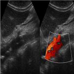
Moving flap is seen in abdominal aorta on real time ultrasonography suggests dissection of abdominal aorta. Aortic dissection occurs when a tear in the inner wall of the aorta causes blood to flow between the layers of the wall of the aorta, forcing the layers apart.
As the patient recently undergone angioplasty, catheter induced aortic dissection is likely.
MRI confirms the sonographic findings. Dissection started from the arch of aorta up to infra renal region.
|